Welcome to the Amira-Avizo Software Use Case Gallery
Below you will find a collection of use cases of our 3D data visualization and analysis software. These use cases include scientific publications, articles, papers, posters, presentations or even videos that show how Amira-Avizo Software is used to address various scientific and industrial research topics.
Use the Domain selector to filter by main application area, and use the Search box to enter keywords related to specific topics you are interested in.
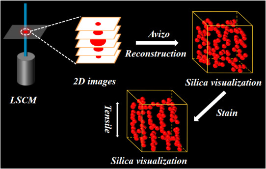
For organic-inorganic composite materials, the spatial dispersion of inorganic fillers in the organic matrix is of great significance for designing and manufacturing high-performance composite materials. To improve the understanding of the micro-physical mechanism of the filler-reinforced polymer matrix, we studied the relationship between filler network structure and macro-mechanical properties of silicone rubber by using fluorescent labeling technology and three-dimensional (3D) visualizati... Read more
Shangjun Zeng, Ming Kang, Kexu Chen, Rong Sun, Ai Lu, Guanjun Changa
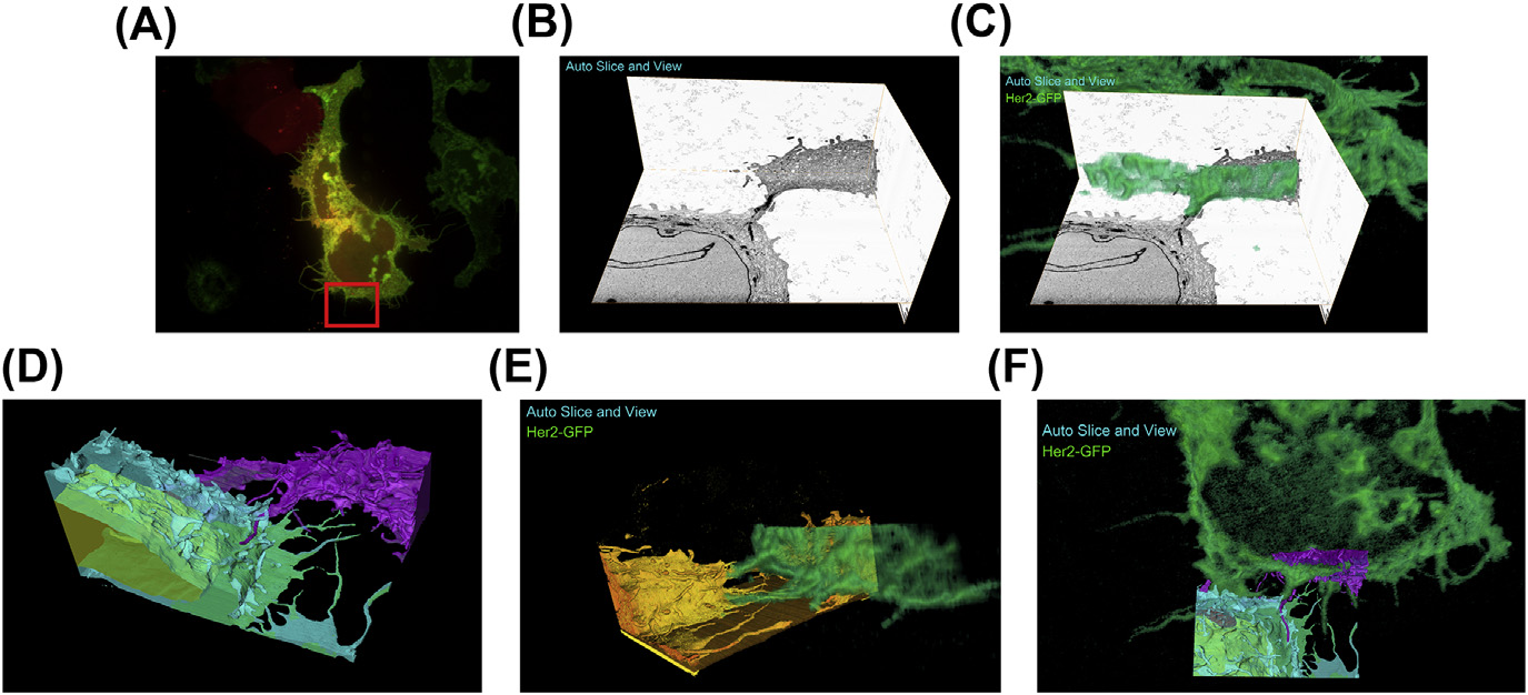
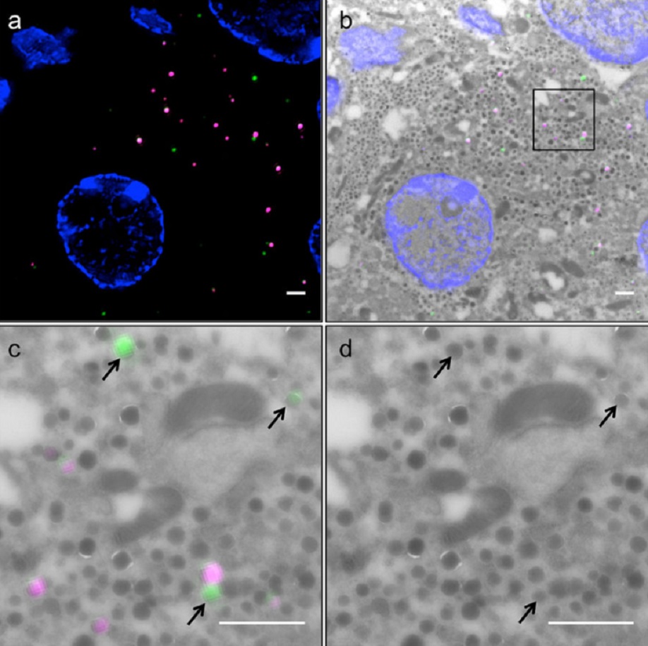
A fully integrated, three-dimensional fluorescence to electron microscopy correlative workflow
While fluorescence microscopy provides tools for highly specific labeling and sensitive detection, its resolution limit and lack of general contrast has hindered studies of cellular structure and protein localization. Recent advances in correlative light and electron microscopy (CLEM), including the fully integrated CLEM workflow instrument, the Thermo Scientific CorrSight with MAPS, have allowed for a more reliable, reproducible, and quicker approach to correlate three-dimensional time-lapse... Read more
Claudia S. Lopez, Cedric Bouchet-Marquis, Christopher P. Arthur, Jessica L. Riesterer, Gregor Heiss, Guillaume Thibault, Lee Pullan, Sunjong Kwon, Joe W. Gray
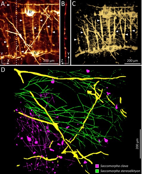
Microscopic organisms that penetrate calcareous structures by actively dissolving the carbonate matrix, namely microendoliths, have an important influence on the breakdown of marine carbonates.
Microscopic organisms that penetrate calcareous structures by actively dissolving the carbonate matrix, namely microendoliths, have an important influence on the breakdown of marine carbonates. The study of these microorganisms and the bioerosion traces they produce is crucial for understanding ... Read more
Philipp-Konrad Schätzle, Max Wisshak, Andreas Bick, André Freiwald, Alexander Kieneke
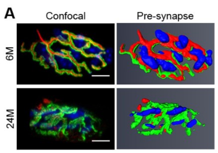
Loss of adult skeletal muscle stem cells drives age-related neuromuscular junction degeneration
Neuromuscular junction degeneration is a prominent aspect of sarcopenia, the age-associated loss of skeletal muscle integrity. Previously, we showed that muscle stem cells activate and contribute to mouse neuromuscular junction regeneration in response to denervation (Liu et al., 2015). Here, we examined gene expression profiles and neuromuscular junction integrity in aged mouse muscles, and unexpectedly found limited denervation despite a high level of degenerated neuromuscular junctions. In... Read more
Wenxuan Liu, Alanna Klose, Sophie Forman, Nicole D Paris, Lan Wei-LaPierre, Mariela Cortés-Lopéz, Aidi Tan, Morgan Flaherty, Pedro Miura, Robert T Dirksen, Joe V Chakkalakal
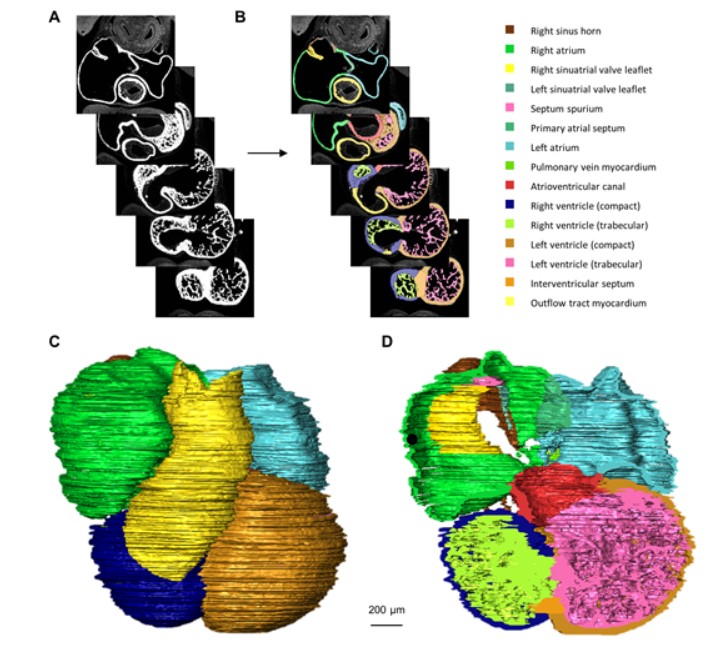
Quantified growth of the human embryonic heart
The size and growth patterns of the components of the human embryonic heart have remained largely undefined.
To provide these data, three-dimensional heart models were generated from immunohistochemically stained sections of ten human embryonic hearts ranging from Carnegie stage 10 to 23. Fifty-eight key structures were annotated and volumetrically assessed. Sizes of the septal foramina and atrioventricular canal opening were also measured. The heart grows exponentially throughout embr... Read more
Jaeike W. Faber, Jaco Hagoort, Antoon F. M. Moorman, Vincent M. Christoffels, Bjarke Jensen
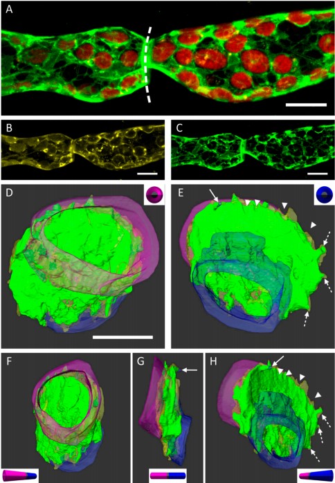
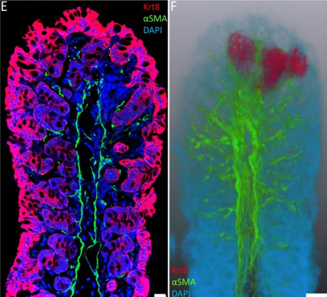
3D Dissection of Structural Membrane-Wall Contacts in Filamentous Moss Protonemata
Cell-to-cell contact is essential for communication and development of multicellular organisms. A prerequisite is the passage through membranes. That way, molecular exchange and information flow is regulated via hormones, membrane proteins and pores.
In plants, the rigid cell walls prevent large membrane contact areas between protoplasts. Only plasmodesmata, minute channels between adjacent cells, form direct connections. Often, molecular data of the proteins involved are manifold but t... Read more
Dominik Harant and Ingeborg Lang
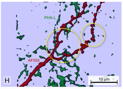
Neuroanatomical tract-tracing techniques that did go viral
Neuroanatomical tracing methods remain fundamental for elucidating the complexity of brain circuits. During the past decades, the technical arsenal at our disposal has been greatly enriched, with a steady supply of fresh arrivals. This paper provides a landscape view of classical and modern tools for tract-tracing purposes. Focus is placed on methods that have gone viral, i.e., became most widespread used and fully reliable.
Read more
Jose L. Lanciego; Floris G. Wouterlood
Analysis of neuronal arborization and connections is a powerful tool in fundamental and clinical neuroscience. Changes in neuronal morphology are central to brain development and plasticity and are associated with numerous diseases. Golgi staining is a classical technique based on a deposition of metal precipitate in a random set of neurons. Despite their versatility, Golgi methods have limitations that largely precluded their use in advanced microscopy. We combined Golgi staining with fluore... Read more
Katlijn Vints, Dorien Vandael, Pieter Baatsen, Benjamin Pavie, Frank Vernaillen, Nikky Corthout, Vasily Rybakin, Sebastian Munck & Natalia V. Gounko
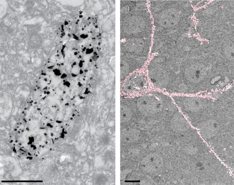
Correlative light and electron microscopy (CLEM) is a powerful approach to investigate the molecular ultrastructure of labeled cell compartments. However, quantitative CLEM studies are rare, mainly due to small sample sizes and the sensitivity of fluorescent proteins to strong fixatives and contrasting reagents for EM. Here, we show that fusion of…
Read more
Andreas Müller, Martin Neukam, Anna Ivanova, Anke Sönmez, Carla Münster, Susanne Kretschmar, Yannis Kalaidzidis, Thomas Kurth, Jean-Marc Verbavatz & Michele Solimena
Immunofluorescence tomography is a high-resolution 3-D reconstruction method based on methacrylate embedding and serial-sectioning, where 2-D images of immuno-stained serial-sections are computationally aligned into image stacks, and the 3-D volume rendered. Butyl-Methyl Methacrylate (BMMA) plastic was adopted as it preserves excellent tissue morphology and can be de-plasticized easily using an organic solvent, which enables immuno-staining of serial-sections without antibody penetration issu... Read more
Parfitt, Geraint J
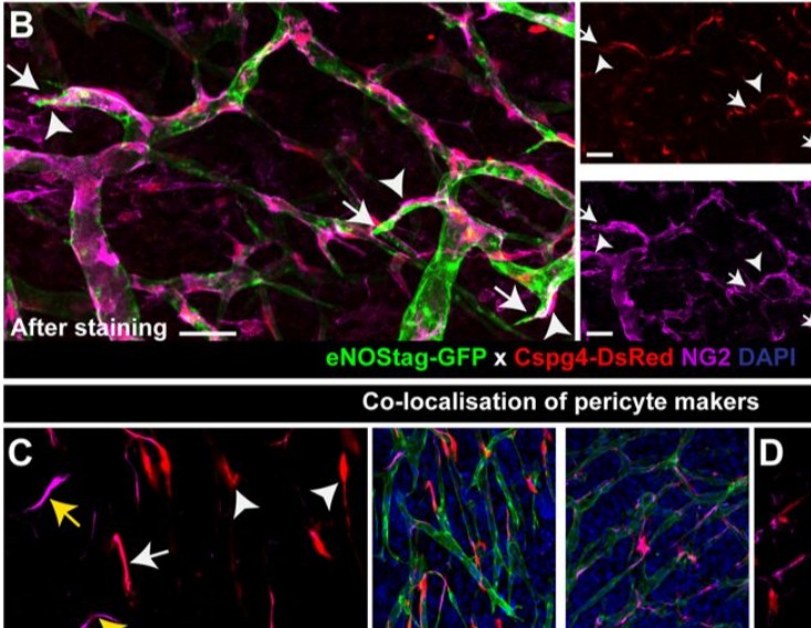
Endothelial cells and pericytes are integral cellular components of the vasculature with distinct interactive functionalities. To study dynamic interactions between these two cells we created two transgenic animal lines. A truncated eNOS (endothelial nitric oxide synthase) construct was used as a GFP tag for endothelial cell evaluation and an inducible Cre-lox recombination, under control of the Pdgfrb (platelet derived growth factor receptor beta) promoter, was created for pericyte assessmen... Read more
Ann L. B. Seynhaeve, Douwe Oostinga, Rien van Haperen, Hanna M. Eilken, Susanne Adams, Ralf H. Adams & Timo L. M. ten Hagen
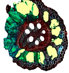
L4iS uses Avizo software to analyze 3D images from laser ablation tomography
Lasers for Innovative Solutions, LLC (L4iS) is developing a new class of tomography technology with the aim of allowing material characterization in three dimensions with sub-micron resolution. The method uses a nanosecond, Q-switched, pulsed ultraviolet laser coupled with high-resolution imaging to generate highly detailed specimen models. Using this system, sequential images similar to light-sheet fluorescence microscopy are used to digitally reconstruct the specimen.
Read more
Brian Reinhardt and Benjamin Hall, L4iS (USA)